


U13-E01. MicroCT GE eXplore Locus SP (GE)
Temporarily OUT OF ORDER

Temporarily OUT OF ORDER
TEMPORARILY OUT OF ORDER
Dynamic mechanical analysis Instron 8874.
UNIAXIAL TENSILE TEST INSTRON MicroTester: Monotonic, Cyclic and relaxation test
Confined compression tests Instron MicroTester Monotonic, Cyclic and relaxation test; Cyclic with relaxation.
U13-S07. Uniaxial tensile nano Bionix MTS monotonic and other mechanical tests
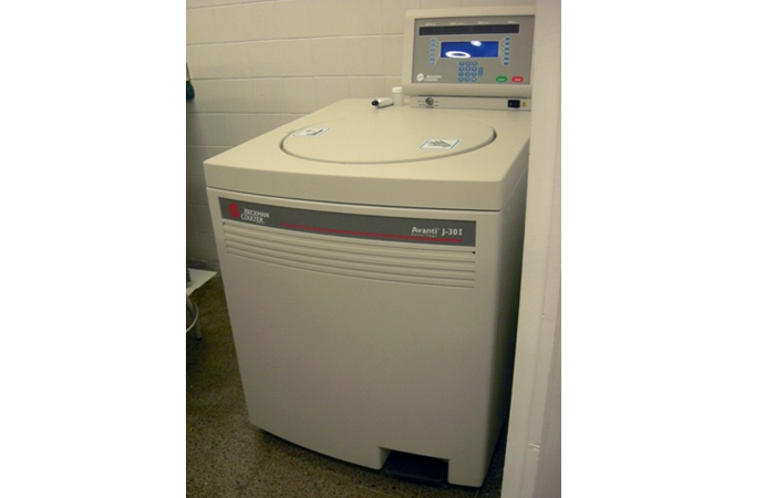
U12-E14. Avanti J30I ultracentrifuge (Beckman Coulter)
Description:
High-speed centrifuge with aluminum rotors at fixed angle – Rotor JA-25.15 (maximum centrifugal force = 74200g, maximum speed = 25000 rpm, capacity for 24 tubes of 15 mL each, total capacity = 360 mL) – Rotor JLA-16.250 (maximum centrifugal force = 38500g, maximum speed = 16000 rpm, capacity for 6 tubes of 250 mL each, total capacity = 1500 mL) – Rotor JA-10 (maximum centrifugal force = 17700g, maximum speed = 10000 rpm, capacity for 6 tubes of 500 mL each, total capacity = 3 L)
Applications:
It allows to centrifuge samples such as emulsions, dispersions, etc at angular velocities up to 25000 rpm (74200 g) at various temperatures (from -20˚C a 40˚C), which can be used for phase separation, stability studies, etc. in colloidal systems and for the separation of cells and proteins.
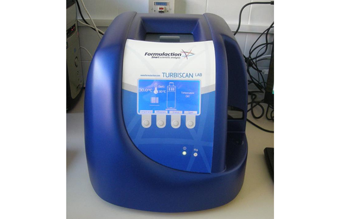
U12-E13. Turbiscan-lab-backscattering-stability-analyzer-formulaction
Description:
Instrument for the evaluation of colloidal stability. It measures turbidity by multiple light scattering, as a function of time along the vertical axis by both transmitted and backscattered light. It can analyse samples up to 95 vol% with a wide range of particle sizes (10 nm – 1 mm). The samples are placed inside a temperature-controlled chamber, and the measurements can be carried out from room temperature up to 60 ˚C.
Applications:
Allows to study the colloidal stability of emulsions, suspensions and foams and to determine whether there is sedimentation, creaming, flocculation or coalescence.
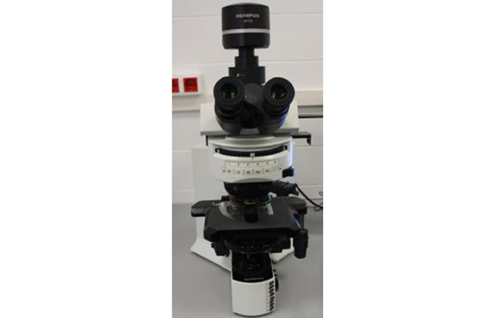
U12-E12. BX51TRF6 Olympus Optical Microscope
Description:
Optical microscope that can work with transmitted or reflected light. Different observation methods can be used: bright field, dark field, phase contrast, differential interference contrast, polarized light, and fluorescence. It includes two revolvers to work at short or long distances. Moreover, it is equipped with a digital camera (Olympus DP73) connected to the computer with a software that captures images and videos.
Applications:
Observation of samples at microscale