+34 9340006100 -ext. 437807-info@nanbiosis.com

Posts on Jan 1970

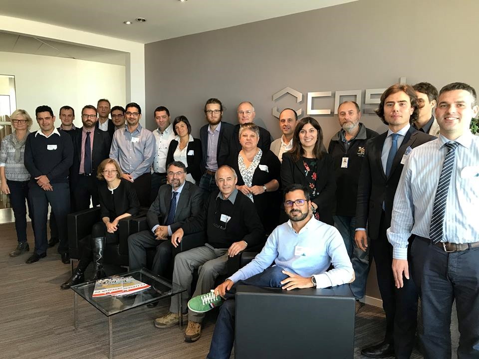
JUMISC, partner of NANBIOSIS, represents Spain in a European network of 22 countries

The Jesús Usón Minimally Invasive Surgery Centre (JUMISC), partner of NANBIOSIS toguether with CIBER-BBN,  leads a multidisciplinary European research network to improve present urinary stents and prevent their causes of failure.

The idea of creating the network was born from a research, started in 1999 by Doctor Soria, Researcher-Coordinator of the Endoscopic Surgery Area at the JUMISC and the promoter of the mentioned network, representing Spain.

His proposal has been supported by the European and Spanish Urology Association, and funded by the Horizon 2020 Program-Cost Actions (European Cooperation in Science and Technology)

The focus of this Action is to create a multidisciplinary team to identify the problems concerning the urinary stents (design, composition, biomaterial applications, coatings, incrustation, etc.) and to promote future research on this field. In this way, the network will try to reduce adverse effects provoked by the present stents.

This European network (2017-2021), coordinated by Dr. Soria, the Chair of the Action, is composed by 22 European countries and other proposals coming from USA,  Japan, South Korea,  Israel, Canada, Russia, India and it is open to other invited countries to cooperate inside  the organised working groups.

This action will provide, during the next 4 years, an extensive and interdisciplinary R&D program to discover the causes of failure of the present stents and will propose cooperative work in urology and bioengineering field.

This proposal, an initiative of the JUMISC, leaded by Doctor Sánchez Margallo, will contribute to improve healthy-quality of patients, reduce in health care costs, and increase the competitiveness of the European medical device industry.
Read More

New Zealand White rabbit as a model for preclinical studies addressing tissue repair at the level of the abdominal wall.

Juan Manuel Bellón and Gemma Pascual, Scientific Director and Coordinator of Unit 17 of NANBIOSIS, have recently published an article in the prestigious journal Tissue Eng Part C Methods.

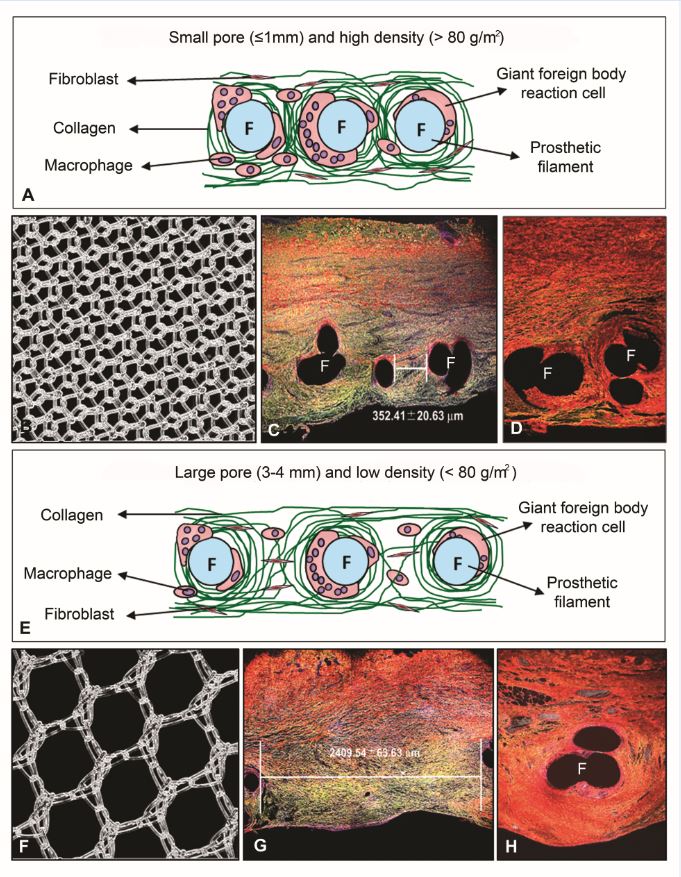
Once a prosthetic material is implanted, an inflammatory reaction is induced in an attempt to remove it or isolate it from the host tissue. After this initial response, a cellular response takes place that gives rise to the start of the process of integration of the biomaterial within the host tissue. Immunohistochemical studies can provide information on the events that regulate this process, and are useful to analyze the progression of the implant and the neoformed tissue. For the tissue repair of a damaged area constantly subjected to tensile forces such as the abdominal wall, optimal collagenization is an extremely important factor. The proper deposition and organization of mainly collagen type I in the neoformed scar tissue will condition the mechanical strength and compliance of the repair zone and thus possible hernia recurrence. In this report, we review the use of the New Zealand White rabbit as the experimental animal for several models of abdominal wall repair and among other techniques, collagen component of the implants was assessed by immunofluorescence using anti-collagen type I and type III monoclonal antibodies. Samples were examined using a confocal microscope Leica SP5 belonging to the platform ICTS ‘NANBIOSIS’ U17 of the CIBER-BBN at the University of Alcalá.

Important information gained from selected models was observed in terms of tissue ingrowth and collagen deposition in the implant area showing significant differences depending on the properties of the implanted prosthetic material. Some variations were also observed in biomechanical properties according to the components comprising each prosthetic material.

Read More