+34 9340006100 -ext. 437807-info@nanbiosis.com

Nanbiosis

U28-E08. FEI Quanta 250 FEG Scanning Electron Microscope + ESEM Mode
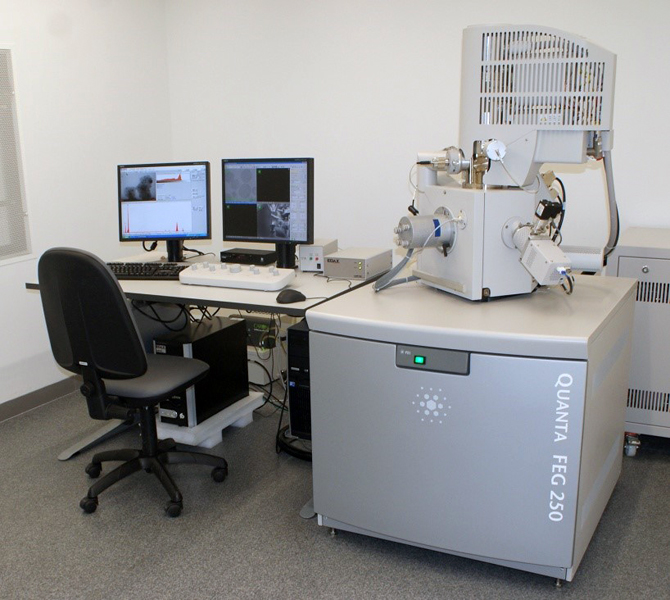
Specifications:
Everhart-Thornley detector of secondary electrons for high vacuum applications providing topographical information
High contrast backscattered electron detector optimized to work with low volt-ages: vCD detector. Suitable for high-vacuum and low vacuum modes, provides topographical and compositional contrast
Scanning-transmission electron microscopy detector (STEM) for high vacuum applications. Allows to introduce 8 grids in one vacuum cycle and to perform bright field and dark field analysis
Large field detector (LFD) for low vacuum and ESEM applications. Allows working in SE and BSE modes and facilitates the observation of large fields of view
Gaseous secondary electrons detector (GSED) for environmental-SEM (ESEM) applications
Gaseous analytical detector (GAD) for ESEM applications
Wet scanning-transmission electron microscopy detector (Wet-STEM) for
ESEM applications

Read More

U28-E07. FEI Tecnai G2 20 TWIN Transmission Electron Microscope + Cryo-TEM

U28-E07. FEI Tecnai G2 20 TWIN Transmission Electron Microscope + Cryo-TEM

Specifications:
Electron source with LaB6 emitter and flexible high tension between 20 and 200 kV
Twin objective lens for high contrast and high tilt applications
Side-mounted CCD Olympus Veleta with 2k x 2k MP, high field of view and fast read-out rates for real time observation
Bottom mounted CCD FEI Eagle, 4k x 4k MP, HS scintillator
EDS thanks to EDAX DPP-II analyzer with Si (Li) detector
Low-dose control software mode for electron beam damage protection of the samples.
Xplore3D software for automated tomography control.
Inspect3D software for post alignment of tomograms.
Amira software for 3D reconstruction.
Sample holders:

  • Single tilt
  • Low background (for EDS applications) double tilt
  • Fischione 2020 advanced tomography holder
  • GATAN Tomography holder for samples at room temperature.
  • GATAN Cryo-station for transferring of cryo-samples to the cryo-holder, previously vitrified of frozen with the high-pressure system.

Read More

U28-E06. Bruker Minispec MQ-60

U28-E06. Bruker Minispec MQ-60

The Minispec TD-NMR is broadly used in research, development and quality control of MRI Contrast Agents. These agents are used to enhance contrast in Magnetic Resonance images between tissues that otherwise would be difficult to differentiate. Most of these contrast agents are based on Gd chelates or iron oxide nanoparticles, which produce signal contrast enhancement by affecting the magnetic relaxation properties of water molecules. The Minispec MQ-60 provides T1 and T2 measurements with high accuracy and reproducibility, and, most importantly, at clinical field, which is fundamental for translational research. Hence, the Minispec MQ-60 plays a key role in the characterization of new contrast agents for clinical use.

Specifications:
Permanent 1.4T magnetic field.
“Intelligent” 1H probe (“plug and play”) with 60 MHz operating frequency
Fast magnet warm-up and precise digital temperature regulation for ultimate results stability
Optical detector to any kind of glass, quartz and plastic samples. Specific filters for natural and fluorescent light suppression and energy saving lamps

Read More

U28-E05. Bruker Avance 600 MHz NMR spectrometer + HR-MAS Probe

U28-E05. Bruker Avance 600 MHz NMR spectrometer + HR-MAS Probe

Specifications:
UltraShieldTM Plus 14.1 T magnet
5 mm BBFOPLUS probe, optimized for X-nuclei direct observation in a frequency range between 31P and 15N. Automated tuning and matching (ATM).
5 mm TXI probe, optimized for higher sensitivity 1H-nuclei observation. The outer coil is optimized for decoupling of 13C and 31P.Automated tuning and matching device (ATM).
TCI CryoProbe Prodigy, uses nitrogen-cooled RF coils and preamplifiers for 1H, 2H and 13C, delivering a 2.5x 1H sensitivity boost. The probe is ATM compatible and has 2H “lock” channel and Z-gradient.
Control temperature unit (from 0 ºC to 50 ºC)

Read More

U28-E04. Bruker Ascend 400 MHz NMR spectrometer

U28-E04. Bruker Ascend 400 MHz NMR spectrometer

Specifications:
UltraShieldTM Plus 9.4T magnet
5 mm BBFOPLUS probe, optimized for X-nuclei direct observation. Broad band in a frequency range between 31P and 15N. This probe has 2H “lock” channel and z gradient that allows 2D spectroscopy and hetero correlation 19F/1H and 1H/19F, to be performed. It also includes an automated tuning and matching device (ATM)
Sample Xpress Lite (for 16 samples)
Control temperature unit (from 0 ºC to 50 ºC)

Read More

U28-E03. Bruker Albira small animal CT system

U28-E03. Bruker Albira small animal CT system

Specifications:
Dedicated beds for individual rats and mice with integrated gas anesteasia.
Albira Software Suite 5.0 with integrated acquisition and tomography software.
PMOD biomedical image quantification software.
X-ray Focal Spot Size (Nominal) 35 μm.
Energy Range (kVp) 10-50.
Field of View (FOV) (transaxial x axial) 70 mm x 70mm
Multiple FOV windows can be combined for whole animal imaging
Bore Size 105 mm

Read More

U28-E02. Bruker In vivo Xtreme

The In-Vivo Xtreme is a multi-modal optical and X-ray small animal imaging system, designed to combine sensitivity, speed and flexibility for the broadest range of applications for molecular imaging. Equipped with a high performance f/1.3 58mm lens and 4MP back-illuminated CCD camera, the In Vivo Xtreme is suitable for demanding bio-luminescence and fluorescence intravital imaging challenges

Specifications:
Back-illuminated 4 MP camera
f/1.1 – f/16, 58 mm fixed lens (on movable platform)
400W Xenon Light Source
28 Excitation Filters 410 nm – 760 nm
Fluorescent imaging from the visible to near IR
Imaging chamber for up to five mice at a time with
integrated anesthesia and temperature control
Automated switching between modalities – the animal never moves
Multimodal Animal Rotation System (MARS)
Integrated high resolution X-ray imaging (>18lp/mm spot size <60μm)
X-ray illumination optimized for small animal imaging with 5 selectable filter settings for fine tuning

Read More

U28-E01. Bruker BioSpec 9.4T animal MRI system

U28-E01. Bruker BioSpec 9.4T animal MRI system


The Biospec 94/20 is a multipurpose research system for high-resolution magnetic resonance spectroscopy and imaging. The system is designed for preclinical and molecular MR imaging. State-of-the-art RF probes technology together with ultra high field USR magnets deliver high spatial resolution in vivo enabling researchers to come closer to the molecular and cellular level (molecular imaging). Thanks to its innovative modular concept, virtually any small animal MR imaging application in life science, biomedical and preclinical research can be conducted.
Specifications:
Magnetic field strength of 9.4T
20 cm bore diameter, suitable for mice and rats
Automated positioning system (AutoPac™)
High performance actively shielded
gradient system with integrated shims
RF coils for rats and mice

Read More

U28-S021. Flow Citometry/Cell Sorter (Onsite&Remote)

Flow cytometry is a highly effective method for analyzing thousands cells or other particles in a matter of minutes. Flow cytometers detect the fluorescence, size and complexity of particles that pass through laser beams as a stream. In this way they can quickly generating statistically valid data by count very large numbers of particles – something that would be extremely difficult with traditional microscopy approaches. Flow cytometers are very well established in hospitals for routine diagnosis and now widely used for different types of research application from marine biology to stem cell research. A Cell Sorter is a specialized flow cytometer which is able to physically separate different classes of particles based on their fluorescence or other physical characteristics.

Applications:
>> Detection of rare events: the high throughput of Gallios allows for the statically-valid detection of very rare cellular events.
>> Multi-parameter assays: Three lasers and ten channels of Gallios allows the design of complex assays using multiple markers.
>> Cell cycle analysis: detection of DNA content, DNA damage, apoptosis and cell cycle antigens and many others.
>> Marine biology: flow cytometry is increasingly used as a tool to study marine microorganisms using their innate fluorescence properties combined with size and complexity characteristics.
>> Single cell separation: Mo-Flo is able to automatically sort single cells into individual wells of a 96-well plate with a very high degree of accuracy and is suitable for colony formation, stem cell isolation etc.
>> Cell purification from tissues: Mo-Flo allows individual cell types from complex tissues to be separated based on size, complexity or markers such as GFP. The yield and purity can be adjusted to suit different applications. Measurement of Relaxation Times (T1, T2, T1*)

Read More

U28-S020. High Content Screening (Onsite&Remote) OUTSTANDING

High content screening (HCS) attempts to bridge the gap between microscopy and cytometry, providing both large quantities of statically useful and phenotypically rich data. High content screening combines automated microscopy with customizable algorithms able to detect and quantify many different types of phenotypic information in both fixed and living cultured cells. Part of the Bionand Cell Culture Unit, the Perkin Elmer Operetta high content screening combines the quality of a spinning disc confocal microscope with Harmony HCS software, which contains many common algorithms such as nuclei and cells detection, as well as the ability to “learn” new phenotypes. Bionand will also offer access to the advanced server-based Columbus HCS software.

Applications:
>> Drug dose response curve calculation: On a single plate multiple concentrations or combinations of compounds can be easily and quickly tested. The software includes all the analysis and presentation tool required to calculate IC50 values. And display response curves
>> Small molecule screens based on changes in cell phenotype: Not all phenotypes can be detected easily by cytometry. Operetta allows very complex phenotypes (e.g. organelle distribution or cell organization changes) to be detected automatically and reliably.
>> Large scale siRNA screens: Automatic detections of up to 384 different siRNAs on a single plate and analysis them automatically makes it possible to perform genome-scale screens for phenotypes of interest
>> Long term cell culture studies: Operetta is equipped with temperature and CO2 control making it suitable for maintaining live cultured cells in optimal conditions during image acquisition

Read More