+34 9340006100 -ext. 437807-info@nanbiosis.com

Nanbiosis

U28-S09. Electron Tomography (Onsite&Remote)

In order to reveal the true nature of many specimens, the third dimension of the sample must be studied. The majority of the TEM techniques record two-dimensional (2D) projections of a 3D structure. However the complexity of the new material and the biological structures highlights the need to develop tools and techniques to explore the morphologies and compositions of materials in three dimensions. An object viewed from many different angles will generate slightly different projections. These images can be recorded and analyzed to create a tomographic rendering of the specimen. The resolution obtained depends of the diameter of the object and the number of projections.

Applications:
Fundamental research in cellular and structural biology for 3D reconstruction studies, and applications involving soft materials that cannot be resin-embedded due to their structural properties and composition.

Read More

U28-S08. Cryo-TEM (Onsite&Remote) OUTSTANDING

Cryo-TEM is the technique of choice for the visualization of biological suspensions and any other sub-micron particles in solution. Cryo-immobilization is performed through plunge freezing where samples are transferred to the cryo-holder and to the microscope at temperatures close to liquid nitrogen. Visualization with Low Dose mode allows beam damage and re-crystallization risks to be minimized.

Applications:
>> Cellular characterization
>> Tissues characterization
>> Biomaterials characterization
>> Macromolecular complexes characterization

Read More

U28-S07. Transmission Electron Microscopy (TEM) (Onsite&Remote)

Conventional TEM provides structural information of samples prepared to electron transparency in the sub-nanometer range. Once prepared, many biological samples, including bacteria, viruses, yeasts, cells, and tissue sections can be characterized using TEM. Moreover, TEM can be used to characterize many other types of material such as substrates or nanoparticles making it a highly versatile technique.

Applications:
>> Cellular characterization
>> Tissues characterization
>> Biomaterials characterization
>> Macromolecular complexes characterization

Read More

U28-S06. Time Domain NMR (TD-NMR) (Onsite&Remote)

NMR relaxometry refers to the measurement of relaxation times, typically T1 and T2, as a function of magnetic field. This is crucial in the development of new contrast agent for MRI.

Applications:
>> Measurement of Relaxation Times (T1, T2, T2*)

Read More

U28-S05. High Resolution Magic Angle Spinning (HR-MAS) NMR (Onsite&Remote)

HR-MAS NMR is based on solid-state NMR, which uses magic-angle spinning at high speeds to average anisotropic interactions, leading to narrower widths. The moderate spin rates used in HR-MAS allows high resolution spectra to be obtained from intact tissue, i.e., without the need of any metabolite extraction procedure. HR-MAS NMR is widely used for metabolomic studies, especially in cancer research, where the metabolic profile (metaboloma) of tumor biopsies can be characterized using just few milligrams of intact tissue (tissue biopsy). HR-MAS can also be used for the structure characterization and elucidation of partially insoluble compounds.

Applications:
>> Metabolomics of intact tissue samples (tumor biopsies, …)
>> Structure Characterization and Elucidation of partially insoluble compounds)

Read More

U28-S04. High Resolution Liquid NMR (Onsite&Remote) OUTSTANDING

Liquid NMR is a fundamental tool both in chemistry and biomedicine research. Its applications to chemistry range from structure elucidation of small molecules to studies of ligand-protein interactions. In biomedicine, the use of liquid NMR has expanded from the classical metabolic studies using enriched stable isotope tracers to the more recent NMR-based metabolomics of biofluids (urine, plasma, CSF).

Applications:
>> Structure verification and elucidation
>> Molecular mobility: determination of conformation in solution
>> Determination of enantiomeric excess
>> Kinetic and temperature studies of reaction mixtures
>> Metabolomics of liquid samples (CSF, plasma, urine and tissue extracts)

Read More

U28-S03. Computed Tomography (CT) (Onsite&Remote)

Computerised Tomography (CT) allows high quality 3D imaging of preclinical animal models based on the reconstruction of 2D X-ray images captured at different angles. Albira uses dedicated beds for rats and mice with incorporated gas anesthesia. Different image capture modes allow users to balance capture time and image quality. The Albira software suite automates the reconstruction process, while the included PMOD software allows users to perform advance analyses and to export data into popular formats such as DICOM. It allows the possibility of combining CT data with fluorescence.

Applications:
>> Tumour imaging and quantification
>> Bone and implantation studies
>> Anatomical CT data is often used in conjunction with other modalities including fluorescence

Read More

U28-S02. Fluorescent and Bioluminescent Optical Imaging (Onsite&Remote) OUTSTANDING

The use of fluorescent and bioluminescent techniques for small animal-based studies into cancer, arthritis and heart disease have grown tremendously in recent years. Part of the Multimodal Imaging Service, Bionand offers the Carestream In-Vivo Xtreme animal imaging system which allows up to five mice to visualized simultaneously using fluorescence and bioluminence methods.

Applications:
>> Tumour and Stem Cell Tracking
>> Monitoring of Drug Delivery and Targeting
>> High throughput Pharmacodynamic studies
>> Imaging and validating new Probes and Biomarkers
>> Bionand offers a range of complimentary services including animal housing facilities, and histology, microscopy and cytometry services

Read More

U28-S01. Magnetic Resonance Imaging (MRI) (Onsite&Remote) OUTSTANDING

Magnetic Resonance Imaging (MRI) is a very versatile technique that offers not only excellent morphological information, but also functional and physiological. Furthermore, important advances during last years in the development of highly sensitive contrast agents using nanotechnology approaches have brought MRI into the molecular imaging arena. Thus, small animal MRI scanners have become a key tool in the development of magnetic nanoparticles for medical diagnosis and therapy.

Applications:
>> Anatomical Studies: tumors, inflammation, Degenerative diseases, etc.
>> Functional Studies: brain perfusion, vascular permeability, in vivo metabolic profiling, etc.
>> Pharmocokinetics and Biodistribution of new Contrast Agents (or any drug magnetically labeled)
>> Monitoring of Drug Delivery and Targeting
>> Follow up of Therapy Response

Read More

STRYKER & NANBIOSIS start a promising line of biomedical research collaboration

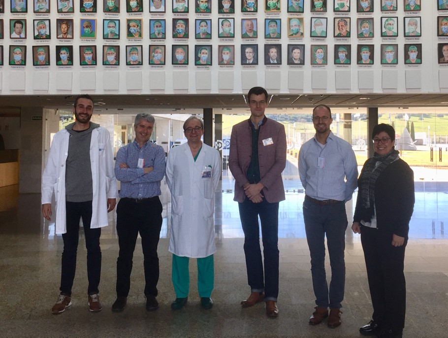
On January 23-24th a two-day meeting between NANBIOSIS and Stryker Trauma GmbH, took place at the Carlos III Health Institute, in Madrid, to explore synergies and potential joint initiatives.  

Nils Reimers and Robin Buescher from Stryker, presented some of their research and upcoming projects in the field of trauma medical devices, and representatives from NANBIOSIS’ units -Marisa González from U16 Surface Characterization and Calorimetry Unit, Teresa Galán from U7, Nanotechnology Unit, Gemma Pascual from U17 Confocal Microscopy Service, from CIBER-BBN, Marisa García from U28 (Bionand) and José Francisco Guillén and Francisco Sanchez Margallo from the units of the Jesús Usón Minimum Invasive Surgery Center (CCMIJU) , together with Jesús, Izco Executive Director of NANBIOSIS and Ramon Martínez, Scientific Director of CIBER-BBN- explained their technologies.

The second day, the meeting location was moved to the CCMIJU in Cáceres, to visit the Center facilities and continue conversations.

All attendees expressed their satisfaction with the development of the meeting and agreed to continue with the explorations in forthcoming meetings.

Photo caption: From left to right: José Francisco Guillén (Quality Guarantee Manager of CCMIJU),  Jesús Izco (NANBIOSIS Executive Director), Francisco Sanchez Margallo (Scientific Director of CCMIJU), Nils Reimers (Senior Manager R&D – Stryker Trauma GmbH), Robin Buescher (Director R&D Stryker Trauma GmbH), Margarita Casado (Management Staff of NANBIOSIS)”

Read More